What is Hernia?
A condition in which part of an organ is displaced and protrudes through the wall of the cavity containing it.
What is Diaphragmatic hernia?
Diaphragmatic hernia (D.H) is an Internal Hernia. Diaphragmatic hernia is the disruption of the diaphragm which allows abdominal organs to migrate into the chest cavity. Diaphragmatic hernia is a disorder which causes great economic loss to dairy industry (Deshpande et al., 1977).
Herniation of the abdominal organs into the thoracic cavity (Bisla et al., 2002).
Hernias of the abdominal wall are common in all domestic species and include umbilical hernias and inguinal or scrotal hernias. Hernias may be direct (through a rent in the body wall) or indirect (through an already existing ring, such as the inguinal ring or umbilical ring).
Diaphragmatic hernia occurs when one or more of abdominal organs enter into the thoracic cavity through a congenital or acquired defect in the diaphragm.
Etiology
- Calving
- Foreign body
- Sudden fall
- Congenital
- Wallowing
Incidence
It is more prevalent in buffalo than other ruminants due to their indiscriminate feeding behaviour as well as the genetic predisposition due to a weak diaphragm.
Clinical signs
- Recurrent Tympany
- Progressive emaciation
- Weakness
- Dehydration
- Regurgitation
Diagnosis:
- History
- Clinical Signs
- Ultrasonography
- Radiography
- Rumenotomy
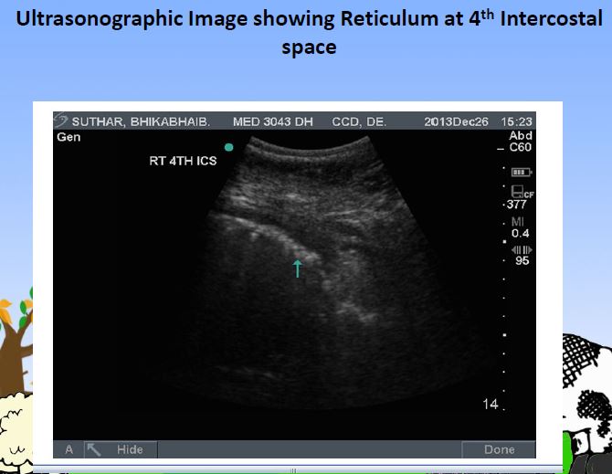
Real time B/M mode Ultrasonography
- Principal: Piezoelectric crystal in transducer sends and receives ultrasound.
- Computer converts the ultrasound echoes to grey dots.
- Each dot correspond to one echo and forms image.
- Anechoic- fluid
- Hypoechoic- soft tissues
- Hyperechoic-air/bone
Restrain and preparation:
- Restrain in standing position in travis
- Shaving hairs at 4th, 5th and 6th intercostal space
- After that application of ultrasound gel
- Technique: 2-5 MHz transducer real time B-mode at 4th, 5th and 6th intercostal space
- Place the transducer vertically in intercostal space
- There is presence of reticular motility at 4th and 5th intercostal space was considered indicative of D.H.


Plain or contrast radiography
- Animal cast in right lateral recumbency
- Plate inserted between sternum and right elbow
- Exposure of diaphragmatic line- juncture of thorax and abdomen
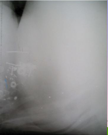
If finds D.H. positive;
- Presence of Foreign Body in thorax
- Diaphragmatic line incomplete
- Animal standing in both legs extended forward
- Exposure of diaphragmatic line: juncture of thorax and abdomen
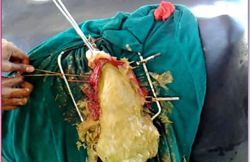
Exploratory rumenotomy
- It is used when other methods fail to diagnose D.H.
- It is also a primary step of D.H. repair
- It decides approach to herniorraphy
- It is used;
- To locate and size of hernial ring
- To remove the foreign body if any
- To access the extent of adhesion
- To evacuate the rumen
Endotracheal intubation:
- Induction:
- Intravenous Diazepam(0.5 mg/kg) + Ketamine(2 mg/kg)
- Intubation:
- Buttler mouth gag
- 18 mm cuffed ET tube
- Digital palpation
- Inflate bulb
- Secure tube
Post-xiphoid trans-abdominal Herniorrhaphy With positive pressure ventillator
- 2 min: Oxygen 4-6 Lit/min
- Isoflurane saturation 5 %
- Deep pain abolish
- Palpebral sluggish
- Isoflurane- maintenance 1-1.5 %

Post-xiphoid trans-abdominal Herniorrhaphy with/without positive pressure ventillator
- Semilateral
- Rig side down
- Laparotomy:
- Post xiphoid curvilinear/ventral midline
Diaphragmatic herniorrhaphy: Incidental Findings/Blunders

TRANS-THORACIC APPROACH
- Incision is made midway between 6th rib and is extended towards costochondral junction.
- 20-25 cm of rib piece is removed by using a rib shear.
- Hernial ring is exposed and remove all the adhesions.
- Now the contents are replaced into the abdominal cavity and sutured in lockstitch manner with braided silk no: 4.
Note: Positive pressure ventilation is needed to be supplied by an endotracheal tube.
Diaphragmatic herniorrhaphy:
- Peritoneum lockstitch pattern chromic catgut no 1
- Muscle sterile silk no 2 – lock stich
- Subcutaneous tissue chromic catgut no 1
- Skin with sterile Silk
- Success rate is 75- 80 %
Source and Credit: Vet NAU Webinar Series 2020 #10 on ”Diagnosis and management of diaphragmatic hernia in bovines” Presented By Dr. Prajwalita Sutaria
Click the below link for the whole presentation with explaination