Pregnancy Diagnosis in Cow
There are various methods for pregnancy diagnosis like:
- Managemental method
- Ultrasound method
- Radiography
- Vaginal biopsy
- Laboratory method
- Clinical method
- Managemental method:
This is based on the history of service by a bull or artificial insemination and non-return to oestrous. This is not a reliable method.
The animal apparently may not return due to various reasons other than pregnancy.
- Ultrasond method:
There are different types of machines available. The most commonly used machines today are B-mode real-time, meaning that they produce an acoustic image in real time.
They usually range from 3.5 – 7.5 MHz. With greater MHz you see more detail but have less depth penetration.
There is more depth penetration with lower MHz, but less detail.
- Laboratory method:
The blood or milk progesterone level is used as an indicator of pregnancy in this method. This method has disadvantages like:
- The test is conducted 21 to 24 days after insemination. Even if the cow is not pregnant it will be in Dioestrus phase with corpus luteum actively producing progesterone, which can give a false positive result
- Requires manual dexterity in handling the equipment
- Embryonic Death after 24 days can mislead
- Clinical method:
- This method is most practical and most reliable
- Pregnancy is diagnosed by per-rectal examination of the animal and the anatomical changes in the reproductive organs like ovaries, uterus, uterine artery and palpation of foetus is taken as the indicator of pregnancy.
- Pregnancy diagnosis is an important tool to measure the success of reproductive management of a cattle herd.
- Rectal palpation is probably the most commonly used method for pregnancy diagnosis.
- Palpation is the procedure of feeling the reproductive tract.
- Although the technique of palpation is relatively simple, the use of breeding records greatly increases the accuracy of the diagnosis and speeds up the palpation process.
- Knowledge of when a cow was bred gives the producer some idea as to the stage of pregnancy, but only if the cow conceived.
- Most producers consider rectal palpation to be the fastest and most accurate method to diagnose pregnancy in cattle.
Equipment Necessary for Palpation
The following equipment is needed to safely palpate a cow:
- Protective covering for palpator,
- Lubricant
Protective Covering for Palpator
- Because the palpator must insert the hand and arm into the cow’s rectum, it is necessary to cover those body parts.
- Disposable plastic sleeves are used for that purpose.
- It is also recommended that the palpator wear protective clothing, such as cover-alls and rubber boots.
Lubricant
A lubricant is applied to the covered hand and arm to facilitate entry into the cow’s rectum.
Commercial obstetrical lubricants are available at farm and ranch supply stores.
A mild liquid soap can also be used as a lubricant, because it provides a slick covering over the arm and does not irritate the cow’s rectal cavity as do some detergents.

Pregnancy diagnosis by rectal palpation remains one of the most practical means for detecting pregnancy in cattle:
Rectal Palpation:
Structures to be palpated
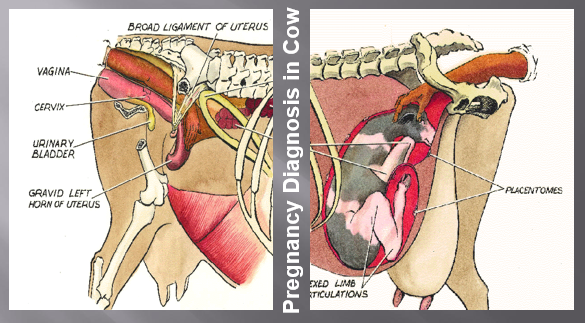
Cervix:
The cervix is chiefly a landmark serving as a guide for locating other structures.
The position of cervix can give an indication of the stage of pregnancy, but a diagnosis should never be based on the cervix alone.
Uterus:
Most of the diagnosis is based on the uterus and its contents.
The size of the uterus (asymmetry) influences its position in relation to the pelvis and should be noted.
The thickness and tone of the uterine wall are important.
The uterine wall becomes thinner as pregnancy progresses and is very resilient to touch compared with the uterus of the open cow.
As pregnancy advances, the cervix and uterus move down over the pelvic ridge and into the body cavity.
Foetal membrane Slip:
Gently grasping the uterine wall between the thumb and forefinger and lifting slightly can detect the chorionic membrane.
With some practice one can feel the membrane slip from between the thumb and finger.
Therefore, the term “slipping of the foetal membrane” has been used to describe this procedure.
By 120 days (4 months) the placentomes are large enough to palpate through the uterine wall.
The contents of the uterus are the most positive diagnostic structures to be palpated.
After 90 days, the foetus can be palpated except during a period from 170-230 days (6-7 months) when it is too deep in the abdominal cavity to reach in large cows.
Amniotic Vesicle
From approximately 30 to 65 days gestation, the amniotic vesicle can be detected as a movable oval object within the uterine lumen..
The vesicle is turgid early in pregnancy but becomes flaccid with advancing pregnancy until days 65 to 70 when it is difficult to detect at all.
The amniotic vesicle can first be palpated at 35 days when it is 7.5 mm in diameter; at 42 days the diameter is 15 mm, at 48 days 35 mm, at 52 days 55 mm, at 58 days 75 mm, at 62 days 90 mm, and at 65 days 105 mm. Initially the vesicle is turgid.
After 60 days the vesicle becomes softer which allows recognition of the small fetus directly.
Placentomes:
The presence of placentomes is another positive sign of pregnancy and is detectable from about 75 days to term.
Since there is great variation in size among individual placentomes, usefulness in aging a pregnancy is limited.
In general, they can be detected as soft, thickened lumps in the uterine wall and are more easily detached as pregnancy advances.
Palpation of the Foetus:
Of course, the presence of the foetus itself is a sign of pregnancy.
Depending on the skill of the examiner and the location of the foetus, the foetus can be palpated from the time of amniotic softening (65 to 70 days) to term.
Foetal growth is quite uniform up to about the sixth month, so that foetal size can be used to estimate foetal age accurately.
Ovaries:
The ovaries can be palpated up to about 120 days.
Structures on the ovary can help confirm either a positive or a negative diagnosis..
Pregnancy is always accompanied by corpus luteum
However, one must remember that a corpus luteum is not always accompanied by pregnancy.
Pulse of pregnancy (Fremitus)
- Helpful in confirming a diagnosis and also confirming the viability of calf, particularly at certain stages of pregnancy
- Felt in the middle uterine artery, which supplies blood to foetus
- By 120 days of pregnancy the middle uterine artery will have enlarged sufficiently to be used as a differential diagnosis in pregnancy determination by rectal palpation.
- Enlargement of the uterine artery ipsilateral to the pregnant horn is detectable after 80 to 90 days of gestation
- By approximately 130 days, the blood flow within the ipsilateral uterine artery has increased to the point at which turbulence is palpable as a buzzing sensation, also referred to as a thrill or fremitus.
Palpation at 35 to 40 days:
The following features should be identified:
- Uterus on the floor of the pelvis, except in large cows with elongated reproductive tracts. Slight enlargement of one horn with detectable dorsal bulging.
- Foetal membrane slip
- Palpation of corpus luteum on the ovary adjacent to gravid horn
Palpation at 45 to 50 days:
- Foetal membrane slip
- Uterus still on pelvic floor. Slightly greater difference in size
- Palpation of corpus luteum on the ovary adjacent to gravid horn.
Palpation at 60 days: (2 months)
- The gravid uterine horn will be dropping slightly over the brim of the pelvis and feels like balloon filled water.
- Foetal membrane slip
- Corpus luteum on the ovary adjacent to gravid horn.
Palpation at 90 days: (3 months)
- The uterus will be pulled well over the pelvic brim and will be 8 to 10 cm in diameter
- The foetus will be 10 to 15 cm long and easily palpated.
- Corpus luteum on the ovary adjacent to gravid horn.
Palpation at 120 days: (4 months)
- The uterus will be well over the brim of the pelvis with the cervix pulled almost to the pelvic brim.
- The foetus can be easily palpated and will be from 25 to 30 cm long.
- Small palcentomes can be identified
- The ovaries may be difficult to reach, but a corpus luteum will be present on the ovary adjacent to the gravid horn.
Palpation at 150 days: (5 months)
- The uterus will be pulled well into the abdominal cavity and the cervix will be located at the brim of the pelvis
- Distinct placentomes about the size of ovaries can be identified.
Palpation at 150 days: (5 months)
- The foetus is well formed and will be 35 to 40cm in length but may be difficult to reach in larger cows.
- The pulse of pregnancy (fremitus) will be quite distinct with the artery being 6 mm to 1.25 cm in diameter.
Palpation at 170 to 230 days (5.5 to 7.5 months)
- Cervix will be at the brim of the pelvis and may be bent over the edge.
- The dorsal wall of the uterus will be tight and difficult to palpate.
- The placentomes will vary in size and may be difficult to palpate because of the tight uterine wall.
Palpation at 170 to 230 days (5.5 to 7.5 months)
- The foetus will be large enough to extend back within range of the hand. The head and front feet are usually the structures palpated.
- Movement of the foetus can frequently be detected.
For more details download PDF and PPT from the attachment.

